


Dr Hofmeyr Viljoen, a Neuroradiologist from SCP Radiology, explains strokes – a critical health issue affecting people of all ages – from its causes to the cutting-edge imaging that guides life-saving care.
In South Africa and across sub-Saharan Africa, stroke rates are rising and it’s a leading cause of adult mortality.
What is a stroke?
It’s essentially a ‘brain attack’ – the brain’s equivalent of a heart attack. It occurs when the blood supply to a part of the brain is suddenly cut off or significantly reduced. Without this vital oxygen and nutrient supply, brain cells in the affected area begin to die within minutes.
This is caused by either:
- A blockage, usually a blood clot, in an artery supplying a part of the brain (Ischemic stroke)
- When a blood vessel in the brain ruptures (Haemorrhagic stroke). The blood can damage brain tissue and result in a dangerous increase in pressure in the skull. It usually presents with a sudden, severe headache and associated weakness on one side of the body
What are the warning signs of a stroke?
When it comes to a stroke, you need to act FAST, an acronym used to recognise the most common warning signs:
F = Face drooping: One side may droop or feel numb.
A = Arm weakness: One arm may be weak or numb.
S = Speech difficulty: Speech may be slurred or the person might be unable to speak
or understand.
T = Time to call emergency services. If you observe any of these signs, call for emergency help immediately.
What warning signs should everyone, especially young adults, be vigilant about?
The warning signs are the same, regardless of age.
Other critical symptoms to watch for include: Sudden numbness, confusion, vision problems, dizziness and loss of balance. These require immediate medical attention.
Is the view that strokes only happen to older people incorrect?
Yes, it’s a common misconception. While strokes are statistically less common in people under the age of 50, when they do occur they can be even more devastating, robbing young people of decades of healthy life. The personal, family and societal costs are enormous.
So, what causes strokes?
The common risk factors – high blood pressure and clogged arteries caused by smoking, diabetes, high cholesterol and family history – account for only 15-25% of cases of strokes n younger people. Other, less typical causes include:
- Blood clots: About 20% of strokes happen when a blood clot travels to the brain, usually originating in the heart
- Blood clots from the rest of the body, such as deep venous thrombosis, are usually filtered by the lungs. However, in patients with small congenital heart defects clots can bypass the lungs and into the brain
- Artery tears: Sometimes, a small tear can form in a blood vessel in the neck or head, often after a minor injury or sudden neck manipulation. This causes up to 20% of strokes in young people
- Blood clotting disorders: Some people have genetic diseases that make their blood more likely to clot
- Inflammation or infection of blood vessels such as HIV and TB
- Drug use: Stimulants like cocaine or methamphetamine are well known causes of strokes in young patients, causing blood vessel inflammation and spasm
- Pregnancy and the first few weeks after giving birth, oral contraceptive use and cancers, are all associated with increased clotting risk
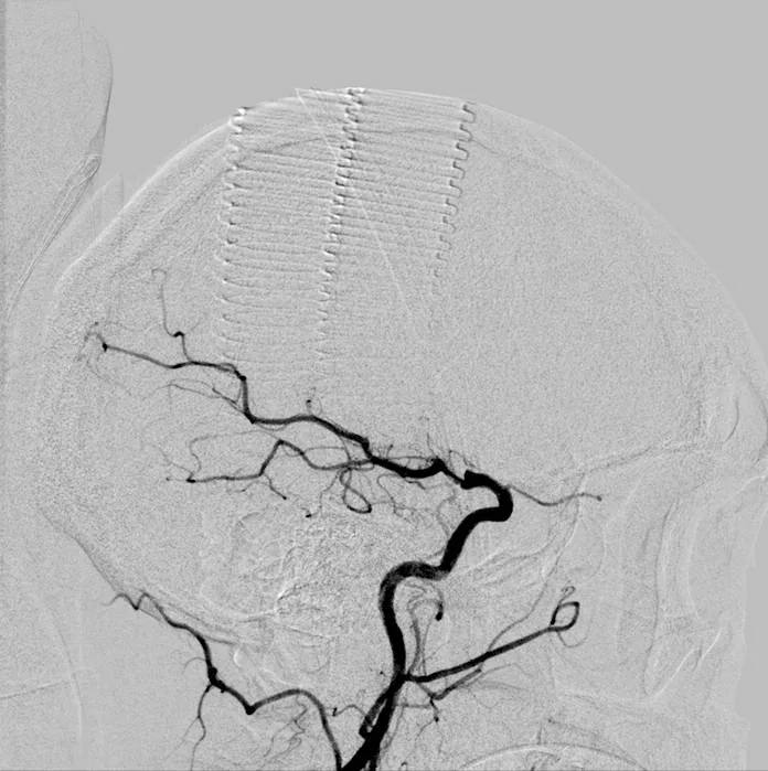
What is radiology’s role in diagnosing a stroke?
Neuroimaging or imaging of the brain is most important for a fast, accurate diagnosis. Our role is to determine if it’s a blockage or a rupture, assess the extent of brain damage and determine what tissue can be salvaged.
What imaging does radiology use in the case of a suspected stroke?
Imaging is the radiologist’s toolkit. We interpret scans rapidly to confirm stroke type, measure extent, identify the problem artery and flag complications. The better the neuroimaging access, the better the outcomes and timely treatments.
- Non-contrast CT of the head: This is fast, widely available and excellent for detecting a haemorrhage
- CT Angiography (CTA) is often performed if no bleed is detected. Contrast (iodine-based dye) is injected into an arm vein during the scan and circulates through the body. It highlights the head and neck blood vessels, pinpointing clots, vessel narrowing, bleeding and tears
- MRI is very accurate in detecting stroke. However, this is less commonly used in the acute setting due to the much longer scan times and limited availability after hours
- Digital Subtraction Angiography (DSA) is a more invasive angiogram where the contrast is directly injected into the neck arteries
What proactive steps can young people take to reduce stroke risk?
Up to 80% of strokes are preventable.
- Know and control your numbers. Regularly check and manage blood pressure, blood sugar and cholesterol
- Manage infections. Get tested for HIV and TB. If positive, adhere to the treatment protocols. Effective HIV therapy reduces associated stroke risk
- Embrace a healthy lifestyle. Don’t smoke, avoid or limit alcohol intake, avoid illicit stimulant drugs and maintain a healthy weight through balanced diet and regular physical activity
Your take home message?
Strokes are not exclusively an ‘old person’s disease.’ Young adults are also at risk. Understanding modern imaging’s power in diagnosis. Know and recognise symptoms, act F.A.S.T. and call emergency services immediately if you suspect you or someone is having a stroke.


